Products
FIB-SEM
Nanomanipulators
OmniProbeOmniProbe CryoSoftware
AZtec3DAZtecFeatureAZtec LayerProbeTEM
Hardware
EDSUltim MaxXploreImaging
TEM CamerasSoftware
AZtecTEM
 Part of the Oxford Instruments Group
Webinar: Watch on demand
In this webcast, the speakers will focus on the biomedical applications of EDS. In this illustration of EDS capability, they will discuss how it can be used to identify the composition of wear particles from failed hip implants and thus determine which part of the implant they originated from.
They will also describe case studies where EDS has been used for medical diagnostics. Finally, they will show how EDS can be used to conduct colour electron microscopy, providing imaging information that facilitates the identification of cells and tissues within hydroxyapatite implants and how the composition of the implant changes during degradation.


Ultimate spatial resolution and low energy performance for EDS in the SEM. Combining Extreme electronics and windowless construction with optimised geometry and sensor design delivers up to 15x greater sensitivity than a conventional large area SDD.
Find out more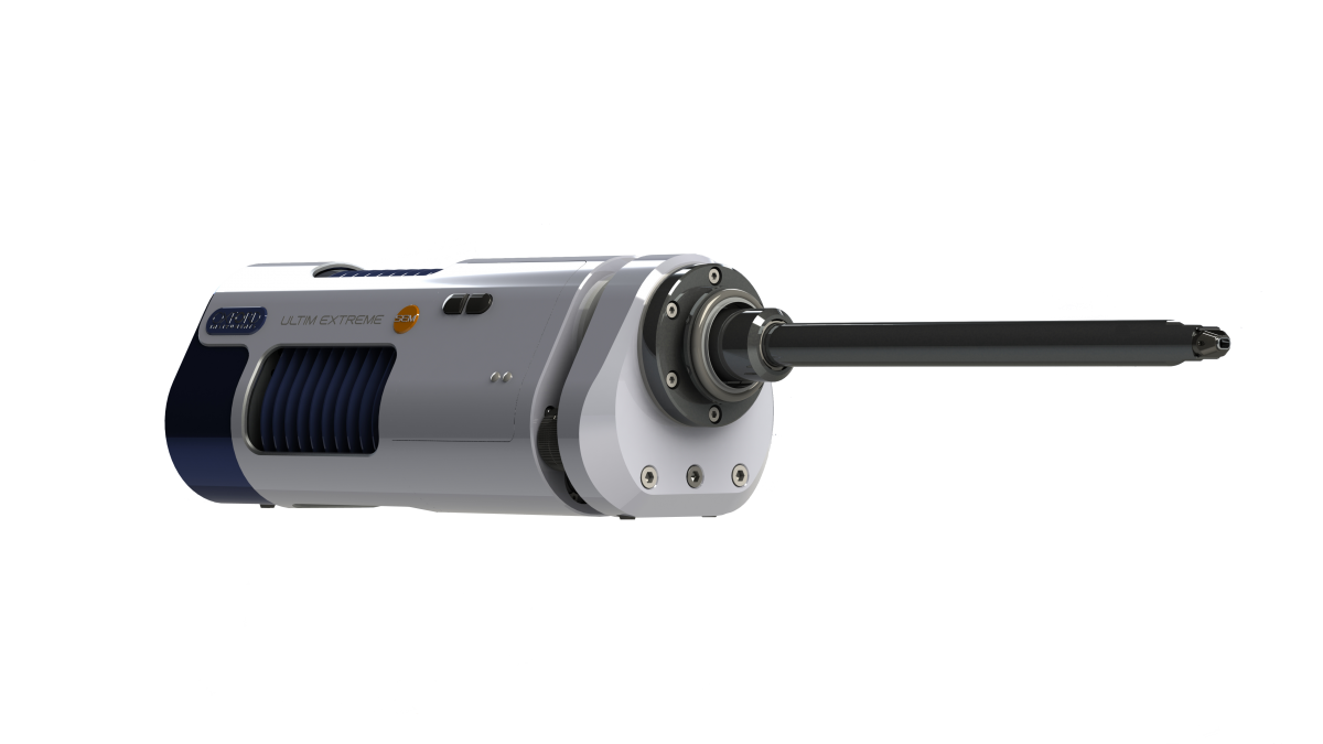
© Oxford Instruments 2024