Fluorescence microscopy relies on precise optical control of excitation and emission light. Excitation filters,
dichroic mirrors and emission filters work together to ensure that fluorophores are efficiently excited and that only
the desired fluorescence signals reach the detector.
This article provides an overview of filter and dichroic mirror configurations commonly available in benchtop
fluorescence microscopes, highlighting the advantages and limitations of different approaches.
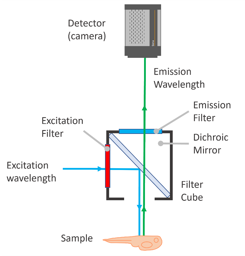
Fig.1 Schematic illustration of the light path in fluorescence microscopy. The excitation filter, dichroic mirror
and emission filter work together to direct selected excitation wavelengths to the sample and selectively transmit
fluorescence emission wavelengths to the detector.
Which Filter and Dichroic Configuration Should I Consider when Choosing a Fluorescence Microscope?
Most benchtop fluorescence microscopes are delivered with preconfigured illumination, dichroic mirrors, and filter
sets optimised for common fluorophores. However, it is essential to verify that the provided configuration is
compatible with your specific fluorophores and experimental requirements.
Excitation Filters
Excitation filters are optical components used in fluorescence microscopes to select the specific band of wavelengths
required to excite fluorophores in a sample. Their role is to ensure that only the desired excitation light reaches
the specimen, while unwanted wavelengths are blocked.
Excitation filters are essential for shaping the relatively broad spectra of light produced in LED-illuminated
widefield fluorescence microscopes. In contrast, laser-based illumination as used in confocal systems, does not
require excitation filters, since lasers provide single-wavelength excitation by default.
| Filter Type |
Description |
| Single-band excitation filters |
- Transmit one narrow excitation band matched to a single fluorophore
- Provide high spectral specificity with minimal bleed-through between channels
- Deliver better signal-to-noise ratios and cleaner images than multiband filters
Disadvantages: - Require separate filters or cubes for each fluorophore
- Slower and less flexible for fast
multi-colour imaging workflows
|
| Multiband excitation filters |
- Contain two or more distinct excitation passbands
- Enable excitation of multiple
fluorophores simultaneously (e.g. DAPI, FITC, TRITC)
- Must be used in combination with matching multiband
dichroic mirrors and emission filters
- Allow faster acquisition when imaging multiple fluorescence channels
Disadvantages: - Increased risk for signal bleed-through between channels
- Higher likelihood of photobleaching
due to simultaneous multi-wavelength excitation
|
Dichroic Mirrors
Dichroic mirrors are optical components placed in the detector beam path that spectrally separate excitation light
from fluorescence emission.
Used in combination with emission filters, dichroic mirrors ensure that only the desired fluorescence wavelengths
reach the detector while reflected excitation light is efficiently rejected. The choice of dichroic configuration has
a substantial impact on which fluorophores can be imaged. Choosing the right dichroic configuration is therefore
essential for accurate and efficient single colour or multi-colour fluorescence imaging.
| Mirror Type |
Description |
| Single-band dichroic mirrors |
- Transmit a single emission band
- Typically used in entry-level benchtop systems
optimised for one or two common fluorophore combinations
- Cost-effective and well-suited for low-complexity,
single-colour or dual-colour assays
|
| Multi-band dichroic mirrors |
- Allow multiple emission bands to be transmitted through a single dichroic mirror
- Enable fast multi-channel imaging without the need for mechanical switching
- When paired with appropriate
excitation and emission filter sets, they provide excellent performance with minimal signal crosstalk
- Preferred choice for systems designed for high-content screening or workflows requiring fast multi-colour
acquisition
|
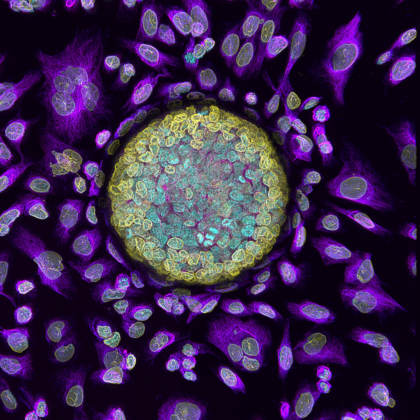
Fig.2 - Multi-colour fluorescence image of a pancreatic organoid. Organoid is mounted in hydrogel and imaged with
BC43. Cyan: DNA; yellow: laminin A/C; magenta: tubulin. Image credits: Dr. Sebastian Amos and Dr. Yu-Suk Choi,
University of Western Australia.
Emission Filters
Emission filters determine which wavelengths of light ultimately reach the detector and therefore play a central role
in defining fluorescence channels and ensuring signal specificity. In practice, these filters transmit the
fluorescence emitted by the fluorophore of interest while blocking residual excitation light and unwanted emission
wavelengths.
The choice of emission filters directly affects image quality and the ability to separate signals in single- and
multi-colour imaging experiments.
| Filter Type |
Description |
| Single-band-pass filters |
- Transmit a single, well-defined emission band - Minimise spectral bleed-through between
channels
- Reduce or eliminate the need for post-processing spectral unmixing
- Ideal for low-light,
high-sensitivity imaging applications
|
| Multi-band-pass filters |
- Transmit multiple emission bands simultaneously
- Allow acquisition of several
fluorescence channels without filter switching
- Improve acquisition speed in time-sensitive or large-area
workflows
- Increase risk for channel crosstalk, requiring careful validation using single-label controls or
post-processing spectral unmixing
|
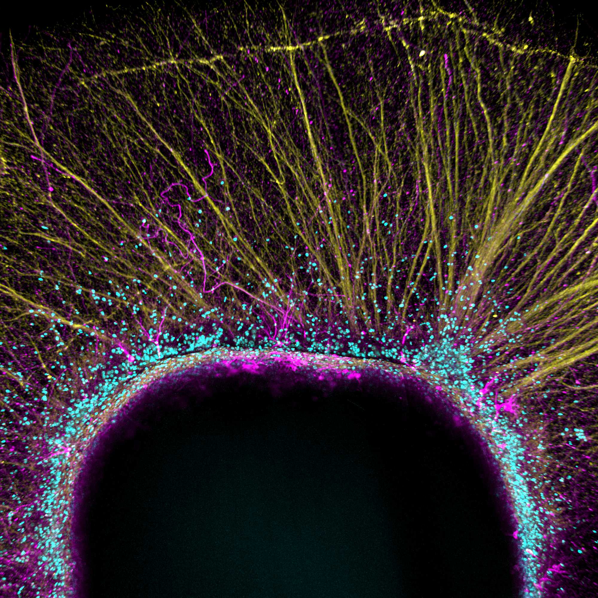
Fig.3 - BC43 multicolour imaging of a mouse embryonic stem cell. Derived dopaminergic neurons cultured on top of
a collagen hydrogel, expressing tyrosine hydroxylase (in magenta),GAP 43 (in yellow), and DNA (in cyan). Image
credit: Ana Marote from ICVS, University of Minho and Leonor Ribeiro from INL
Exploring Benchtop Fluorescence Microscopes?
Learn more about the key technologies, components and practical considerations involved in selecting a benchtop
fluorescence microscope.
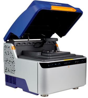
BC43 – Exceptional Performance, Certified Quality, High Productivity
BC43
widefield and confocal imaging systems both use laser illumination and therefore do not require interchangeable
excitation filter sets for wavelength selection.
BC43 incorporates a quad-band dichroic mirror that allows clear band-pass separation of all four integrated laser
lines. This configuration supports rapid multi-colour imaging without the need for mechanical dichroic switching.
BC43 includes single-band emission filters matching the emission wavelengths of fluorophores excited by the four
standard wavelengths used in microscopy (UV, green, red, and far red). Optionally, the system can be equipped with a
quad-band emission filter designed to accelerate live-cell imaging and large multidimensional acquisitions.
← Back to Learning