Breakthroughs and Limitations of Element Mapping
The ability to collect energy dispersive X-ray spectroscopy (EDS) data across a 2D plane revolutionized elemental analysis. This breakthrough allowed the spatial distribution of elements within a solid sample to be visualised—an advancement that transformed materials characterisation. Since its inception, this technique, known as “element mapping”, has not only become exponentially faster but has also revealed unprecedented levels of detail. Today, modern EDS systems can analyse the majority of the periodic table, delivering spatial resolutions from nanometres to centimetres—all within commercially viable timeframes.
Yet, despite these advancements, a fundamental challenge remains; the directional nature of an inclined EDS detector. Even minor surface variations—on the scale of just a few micrometres—can introduce an artifact known as X-ray shadowing. This occurs when elevated features of the sample physically block the emitted X-rays, preventing them from reaching the detector (Figure 1). In essence, the sample casts an X-ray shadow, obscuring critical compositional information from being detected in low areas of topography.
The conventional solution to this problem is straightforward; to cut, grind, and polish the sample until it is completely flat. By doing so, all regions of interest are fully exposed, and shadowing is eliminated.
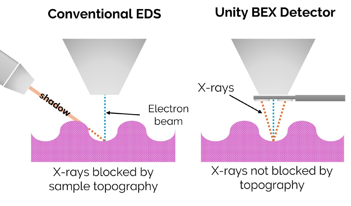
Figure 1: Limitations of inclined EDS detectors when dealing with sample topography by comparison with top-down view of the Unity BEX detector.
While this solution works well for non-precious samples that can be readily modified, it poses a serious dilemma when sample integrity is paramount. What if the material cannot be altered without compromising its structure, value or future use? In such cases, X-ray mapping can become severely compromised, forcing analysts to one dimensional point analysis on topographic samples to overcome low signal.
Greatly Reducing X-ray Shadows with the BEX Technique
The world’s first backscattered electron and X-ray (BEX) detector, Unity, redefines X-ray collection by overcoming a fundamental limitation of conventional EDS detectors. Unlike traditional systems that rely on an insertion angle of approximately 35°, Unity employs two separate sensors beneath the pole piece, with greatly increased solid angle and ‘top down’ view (Figure 1).
This specialised geometry dramatically reduces the impact of X-ray shadowing, a challenge that has long hindered accurate elemental mapping on uneven surfaces. Furthermore, Unity is also equipped with two backscattered electron (BSE) detectors. Permitting simultaneous BSE and X-ray (BEX) acquisition with complete point-by-point data correlation.
As a result, Unity enables high-resolution X-ray mapping where even the most advanced EDS detectors would struggle to produce meaningful data (Figure 1). With this breakthrough, industry and academia alike can now extract compositional insights from complex topographies.
Finding Zircon within One of the Solar System’s Oldest Meteorites
To showcase the applicability of the BEX technique for element mapping on topographic samples, we conducted large-area mapping on an unprepared meteorite. For clarity, the sample was in the exact same state as when it crashed on Earth, with no cutting, polishing or coating. The sample in question was an exceptionally rare extraterrestrial material called ‘angrite’. Angrites are among the oldest known igneous rocks, believed to have originated from the inner solar system approximately 4.35 billion years ago1. Unlocking the secrets within these ancient materials offers invaluable insights into the evolution of the early inner solar system1,2.
Typically, a rock’s geological history is timestamped through the study of radioactive isotopes preserved in the rare accessory mineral zircon. Zircon is a geological time capsule—its uranium-lead (U-Pb) isotopic system evolves at a constant rate from the moment of crystallisation, allowing scientists to date rocks that are millions to billions of years old.
However, retrieving this chronological record is no simple task. In terrestrial rocks similar to angrites, zircon crystals are not only scarce but minuscule. In fact, no zircon has ever been documented within an angrite. For rock samples with sparse zircon, geologists would normally crush and sieve the material to extract and concentrate zircon grains. But with a sample as rare and scientifically valuable as an angrite, destructive methods are not an option. Instead, a non-destructive approach capable of identifying and locating zircon without compromising the integrity of the sample must be found.
Why is BEX the best Approach?
Backscattered electron and X-ray imaging offers an ideal solution to this challenge:
1. Beneath-the-Pole-Piece Insertion - The elevated positioning of the X-ray detectors provides a top-down view of the sample, significantly reducing shadowing effects.
2. Breakthrough Speed - The exceptionally large solid angle of the Unity detector delivers X-ray count rates for Zr that are at least five times higher than those of the fastest EDS detectors. As a result, large-area scans can be completed in just 20% of the time required by a high-end 170 mm² EDS detector. This rapid acquisition is particularly advantageous when searching for ultra-rare phases that may be only tens of micrometres in size.
3. Absolute X-ray and BSE Correlation - These two datasets are acquired simultaneously in the same raster meaning a direct match of this data can be achieved. Therefore, users can be confident in the areas of the sample that the X-ray information correspond to in the event that artefacts like distortion at low magnification occur.
4. Variable Pressure Capability - Unity operates efficiently in variable pressure mode, enabling high-speed analysis of uncoated samples with greatly improved data quality.
5. Peak Deconvolution with TruMap - Peak overlap occurs when an X-ray peak generated by a single element is falsely identified as being derived from another element with similar X-ray energy. Overlap is particularly common when mapping, as high throughput and short process times lead to broader spectrum peaks. TruMap effectively resolves the spectral overlap, applying our leading peak deconvolution algorithms within Tru-Q© IQ to every pixel (Figure 2). This is especially critical when analysing angrites, as the P Kα X-ray produced by apatite could be falsely represented as the Zr Lα X-ray produced by zircon (Figure 2).
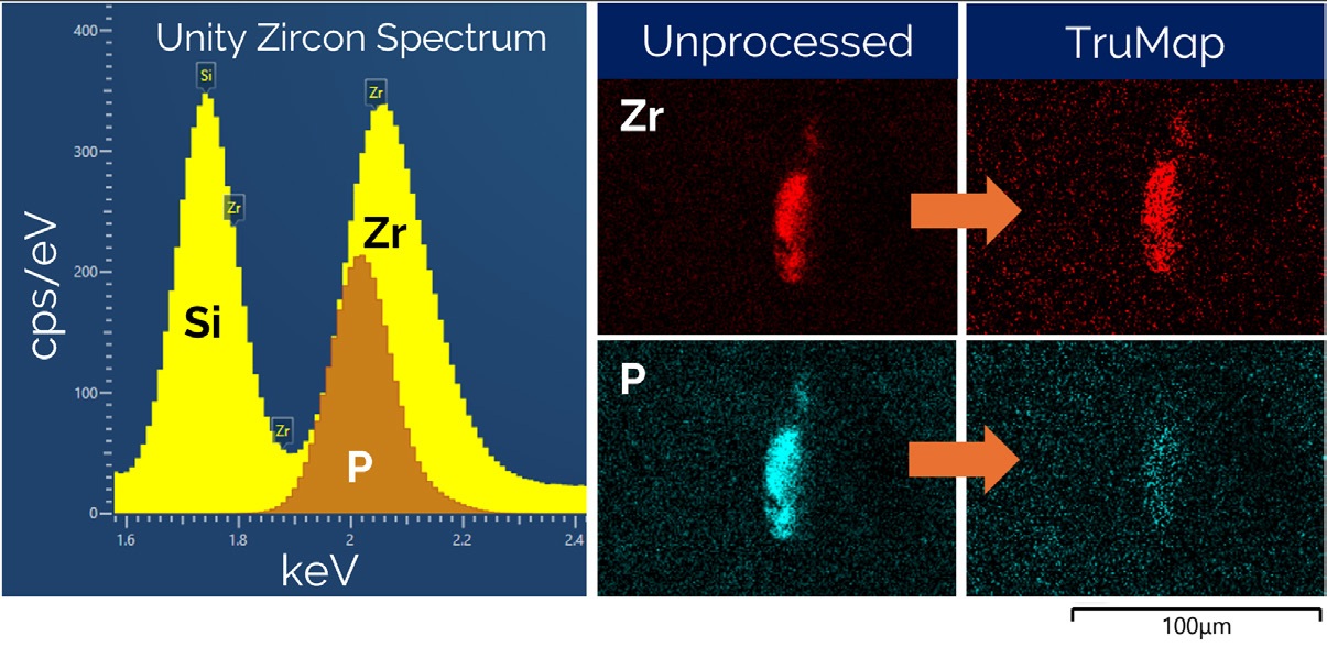
Figure 2: Unity zircon X-ray spectra demonstrating closeness of P and Zr peaks. Images demonstrating the vitality of TruMap’s peak deconvolution when dealing with Zr and P X-rays during high-speed BEX analysis.
Analytical Methodology
Analysis was performed using a Zeiss Gemini 460 scanning electron microscope, equipped with an Oxford Instruments Unity BEX detector and a 100 mm² Ultim Max EDS detector. This way, Unity can facilitate the high-speed X-ray mapping on challenging topography whilst the EDS detector performs element identification with its higher spectral resolution. All measurements were conducted at an accelerating voltage of 15 kV, with a beam current of 4 nA at a working distance of 8.5 mm and a low magnification of 400 X. Variable pressure mode was utilized, maintaining a vacuum pressure of 35 Pa to mitigate surface charging effects.
At the same time, Unity detected 580% more X-ray counts than the 100 mm2 Ultim max ∞ detector. To achieve a similar number of counts with the large area 100 mm2 Ultim max ∞ detector would take 23 hours. Using a standard EDS sensor size of 60 mm2 or less would take at least 38 hours, in addition to significant information loss due to shadowing (Figure 4).
The Zr X-ray TruMap was analysed in isolation to highlight regions of high Zr intensity (Figure 5). As a pixel size of 2.5 μm2 was used, BEX images could be collected at 400 X magnification whilst still being able to resolve very small phases. Low magnification and high resolution was critical for achieving high-speed analysis, being able to cover a large sample within a reasonable timeframe.
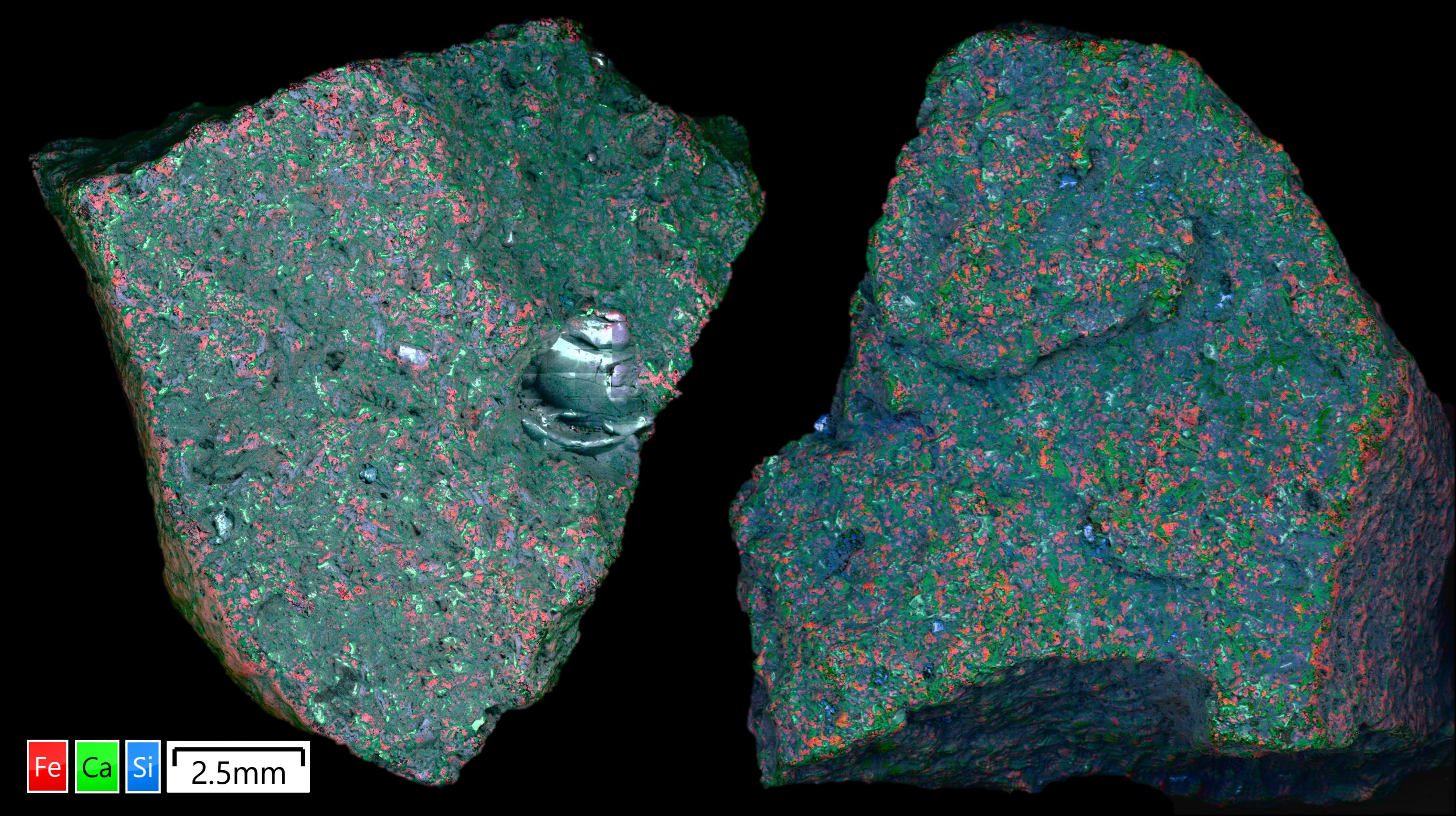
Figure 3: Large area BEX cartography of angrite sample displaying strong X-ray signal across undulating sample surface. Cartography is generated by montaging a series of individually acquired images collected during an automated run.
Results
A total area of 4.5 cm² was mapped using BEX over four hours (Figure 3), generating two high-resolution montage images representing both sides of the angrite. The two maps included 1400 individual fields with a magnification of 400 X and a pixel size of 2.5 μm2. Importantly, mapping was performed over topographic changes on the order of several millimetres which caused significant shadowing of the EDS detector, not seen in the BEX image (Figure 4).
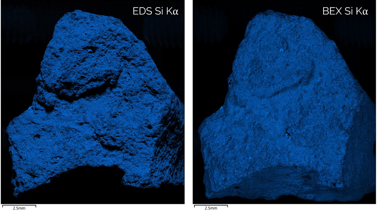
Figure 4: Side by side comparison of Si Kα X-ray intensity across the surface of angrite sample. With zircon sizes less than 50 μm, crystals could easily be lost within X-ray shadows of the EDS dataset.
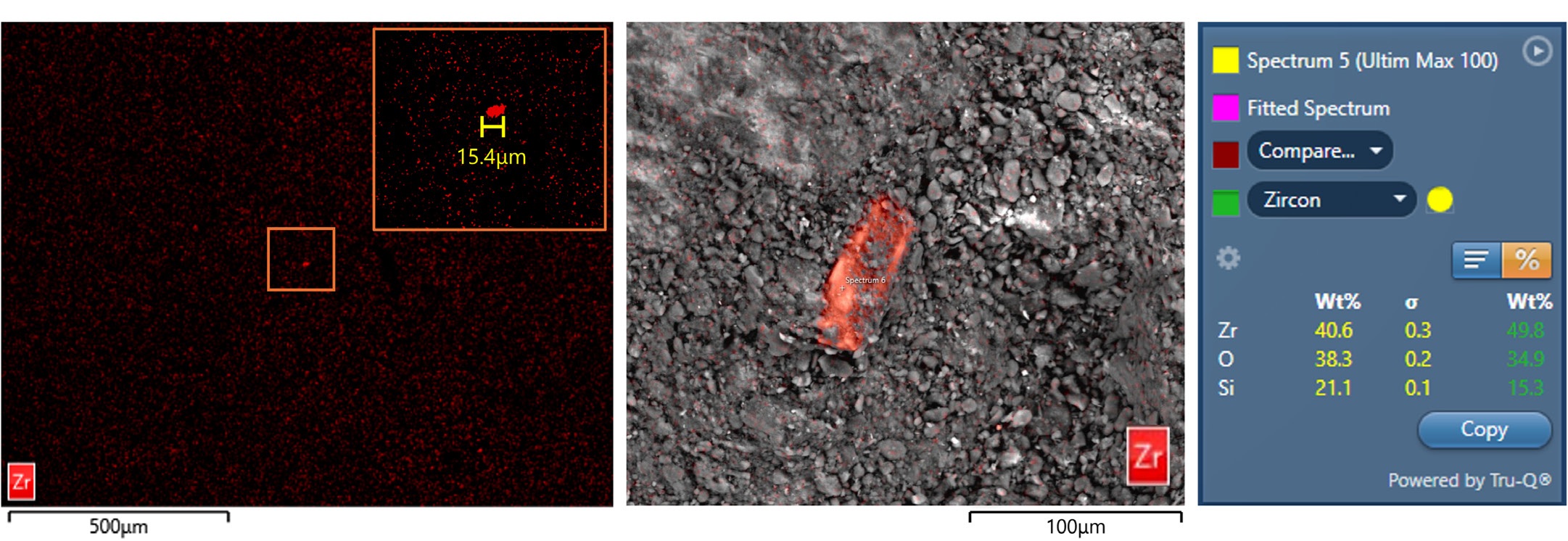
Figure 5: Magnified segment of Zr X-ray TruMap displaying minor zirconium anomalies. High-resolution image and EDS quantification were used to confirm that Zr X-ray peaks represented zircon crystals.
Across both sample surfaces, X-ray mapping successfully identified 10 zircon crystals—marking the first documented occurrence of zircon in angrites (Figure 5). Measuring less than 50 μm in size, these crystals each less than 3 ppm of the total surface area and some were in regions completely obscured from conventional EDS detection due to shadowing. To confirm their composition, several Zr-anomalies positioned outside of shadowed areas were analysed using EDS point analysis, verifying their identity as zircon crystals.
This analysis has led to a groundbreaking discovery in meteoritics—the first detection of zircon in angrite. By leveraging the easy export of BEX maps, this data can now be used to precisely guide focused ion beam (FIB) milling, enabling the extraction of minute zircon-bearing sample volumes. As a result, a significant concentration of zircon crystals can be recovered for U-Pb age dating with minimal sample damage, preserving the integrity of this rare extraterrestrial material.
Conclusion
While conventional EDS mapping is a powerful tool, challenges are found when analysing topographic samples. The BEX method extends the capabilities of X-ray mapping by minimising X-ray shadowing and delivering unprecedented speed with its unique geometry. This breakthrough enables a first-of-its-kind approach for detecting exceptionally rare phases (< ppm abundance) within unprepared samples.
Beyond the potential discovery of the oldest zircons in the solar system - this advancement represents a significant avenue for unravelling the origins of our solar system, paving the way for high-impact research.
Acknowledgements
We would like to thank Dr. Ben Rider-Stokes of the Open University for his collaboration in this study.
References
1. Rider‐Stokes, B.G., Anand, M., White, L.F., Darling, J.R., Tartèse, R., Whitehouse, M.J., Franchi, I., Greenwood, R.C. and Degli‐Alessandrini, G., 2024. The impact history and prolonged magmatism of the angrite parent body. Meteoritics & Planetary Science, 59(1), pp.23-39.
2. Rider-Stokes, B.G., Greenwood, R.C., Anand, M., White, L.F., Franchi, I.A., Debaille, V., Goderis, S., Pittarello, L., Yamaguchi, A., Mikouchi, T. and Claeys, P., 2023. Impact mixing among rocky planetesimals in the early Solar System from angrite oxygen isotopes. Nature Astronomy, 7(7), pp.836-842.

