What is ultrastructure and why is it important?
Ultrastructure is the architecture of cellular material on the sub micrometre scale, and as such is usually imaged using electron microscopy. The cellular structures that have specific functions within cells, known as organelles, exist at the ultrastructural level; these include mitochondria, chloroplast, flagella and granules. Therefore, in order to understand the function and response of cells, it is necessary to image and to identify all structures on the ultrastructural level.
How can EDS assist in ultrastructure classification?
Standard ultrastructural imaging of cellular material necessitates high resolution electron microscopy. This requires complex sample preparation, possibly including staining using heavy metals in order to provide additional contrast in the final images, or specific labelling of antibodies using metal nanoparticles (“immunolabelling”). Interpreting the final images is very challenging: features that have similar morphology, such as granules or immunolabels, can be impossible to distinguish based on image greyscales alone. In these cases chemical information, as provided by EDS, can significantly aid interpretation.
How has Oxford Instruments developed products for the analysis of tissue samples?
Oxford Instruments has developed EDS products that are ideally suited to the analysis of tissue samples. The following developments are key:
- Ultra large area detectors, such as the Ultim Max 170, provide exceptionally high count rates from biological material without requiring high (i.e. damaging) beam currents.
- The pioneering windowless detector, Ultim Extreme, is designed to maximise efficiency at low X-ray energies – precisely where the dominant X-ray signal arises from tissue samples.
- Spectrum processing algorithms ensure optimal performance at the low energy part of the X-ray spectrum, ensuring accurate and reliable results from cellular material.
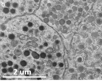
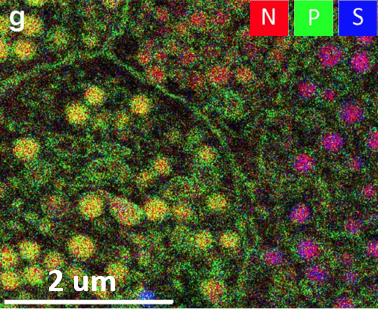
These advances make routine EDS analysis of tissue samples a reality. For example, in research into diabetes, an Oxford Instruments EDS system was used to characterise the granules within pancreatic cells. The SEM and EDS images below show how EDS allows the identification of 2 distinct granules in this cell - zymogen (N rich: red), glucagen (N and P rich: yellow/orange) and insulin (N and S rich: purple). The three granule types would be impossible to distinguish without the use of EDS. These results are taken from M. Scotuzzi et al., Scientific Reports 7, article 45970 (2017) - https://www.nature.com/articles/srep45970.
Back to Bio Imaging & Life Science


