Part of the Oxford Instruments Group
Expand
Bio-Imaging & Life Sciences
Nanoparticle & biomaterials characterisation

Nanoparticle & biomaterials characterisation

EDS for imaging 

Multimodal imaging and the generation of contrast are key research topics when considering the examination of cell and tissue interaction with nanoparticles, biomaterials and implants.  Energy dispersive x-ray spectrometry (EDS) provides imaging contrast using compositional information and combines this with analytical capabilities for measuring the relative quantities of elements within a specimen.

Our EDS systems and analytical software enable rapid and accurate characterisation of samples.

  • Our windowless Ultim Extreme detector is designed for low kV analysis of specimens, providing high resolution mapping data (<2kV) and reducing exposure for beam sensitive and cryo samples.
  • The Ultim Extreme detector is sensitive to the light elements present in biological samples, including nitrogen, resulting in a comprehensive analysis of elements within your specimen.
  • Our EDS systems provide fast and accurate elemental mapping and analysis with large area detectors.
  • AZtec provides easy to interpret analytical data with automatic identification of elements
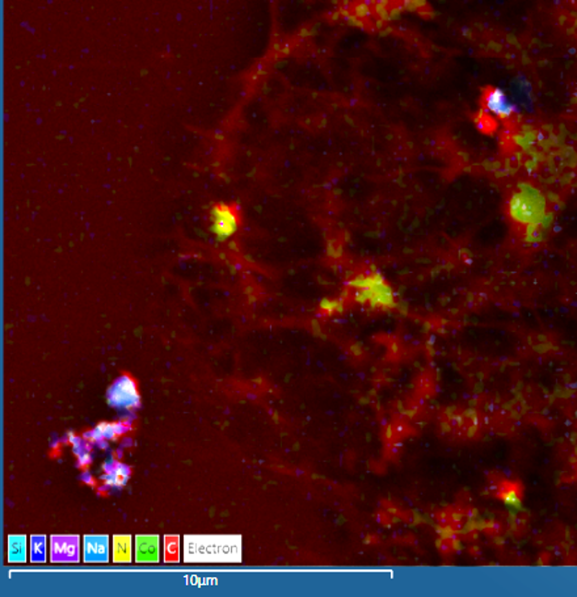
Cultured cells
*Cultured cells containing chromium nanoparticles imaged using an Ultim Max detector in an SEM at 10kV.Sample courtesy of Zhidao Xia, Swansea University 

Nanoparticle Analysis

Wear debris can be produced as a result of friction between adjacent materials potentially causing aseptic loosening of the implant and its subsequent failure. This is particularly evident when the two surfaces are composed of different materials. EDS characterisation of wear debris showed that chromium, titanium and cobalt nanoparticles, originating from different areas on the hip implant, ended up inside the same cells.

BSE image of a TEM section of human tissue
*BSE image of a TEM section of human tissue (left image) showing a cell containing an accumulation of nanoparticles (arrow). Maps of this region (middle and right) reveal that cobalt, titanium and chromium particles are all present in this region of the cell. Data from the human tissue was collected in a SEM using an Ultim Extreme detector at 5kV.  Sample courtesy of Zhidao Xia, Swansea University.

Colour EM for biomaterials

EDS can be used as an imaging tool for colour electron microscopy. EDS provides elemental contrast that can be mapped across a specimen. EDS maps can be used for the identification of features within the sample that are hard to identify using electron data alone, for example the cells (blue & green) shown within the calcium and phosphorous rich implant material that cannot be distinguished easily using backscattered electron images.

*Montages of 240 images showing the overlay of EDS maps onto backscattered electron images. Nitrogen (blue), uranium (green), calcium (yellow) and phosphorous (pink) show the distribution of elements in a biodegradable hydroxyapatite implant, revealing the location of cells and the differentiation between the implant, the mature bone and new bone growth. Sample courtesy of Zhidao Xia, Swansea University.

Library

Webinar: Bringing EDS to Life
Blog: Tips & Tricks for Biological EDS

Key products