Distribution and Identification of Nanoparticles in Cells using EDS
Author: Katarzyna Wulfmeier, Juan Pellico, Alejandra Carbajal, Philip Blower, Vincenzo Abbate, Samantha Terry - Kings College London. Saskia Bakker - University of Warwick. Pedro Machado
Published: 01 Feb 2026 · Last updated: 25 Feb 2026
Tags: EDS Software, EDS
Aim
To confirm the uptake and the subcellular localisation of thallium nanoparticles in cells and tissues for cancer therapy.
Challenge
When investigating the localisation of nanoparticles with electron microscopy, it is necessary to be able to accurately distinguish them from other features within cells and tissues. Biological specimens prepared for electron microscopy are often stained with heavy metals for contrast; therefore, particle electron density may not be enough for definitive identification of nanoparticles.
Solution
The correct identification of the nanoparticles based on their elemental composition was conducted with an Energy Dispersive X-ray Spectrometer Ultim Max TLE mounted on a transmission electron microscope (TEM).
Example
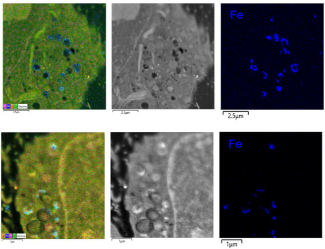
Figure 1. PBNPs were present in vesicular organelles of lung cancer cell lines. Left to right: Electron image and EDS maps overlayed; electron image; Iron (Fe) image maps showing PBNPs.
In this example presented at Microscopy and Microanalysis1, specific cellular uptake of Prussian blue nanoparticles (PBNP) that contain iron (Fe) was investigated in lung cancer cells (Figure 1).
Figure 2. PBNPs can bind thallium (Tl) and its colocalisation can be shown in the image maps. Left to right: Electron image and EDS maps overlayed; electron image; Iron (Fe) image maps showing PBNPs; Thallium (Tl) showing colocalisation with Fe.
In the context of lung cancer, thallium (Tl) can be used to develop a potential radiopharmaceutic therapy. Cells do not normally uptake thallium particles. PBNPs are known to bind to thallium and are therefore a candidate carrier system. We tested this hypothesis using an in vitro cell culture experiment. These particles cannot be accurately identified by electron contrast and size due to other features in the background. Therefore, EDS was used for mapping and comparing the localisation of both PBNPs and Tl (Figure 2).
Conclusion
Combining EDS with TEM offers a solution for elemental analysis combined with ultrastructural information for accurate localisation of nanoparticles. Using EDS, it was possible to distinguish PBNPs from other cellular inclusions and show the binding of thallium.
The ultrastructural and chemical information suggests that the thallium bound to PBNPs localises to secretory pathway organelles (vesicles). In complex experiments where more than one type of nanoparticle is used, EDS elemental maps are essential for the correct interpretation of results.
Acknowledgements
We would like to acknowledge Katarzyna Wulfmeier, Juan Pellico, Alejandra Carbajal, Philip Blower, Vincenzo Abbate, and Samantha Terry for the scientific collaboration that made this application note possible.
References
Subcellular Mapping of Thallium (Tl) Delivered by Prussian Blue Nanoparticles in Lung Cancer Cells — Katarzyna Wulfmeier, Juan Pellico, Pedro Machado, Alejandra Carbajal, Saskia Bakker, Philip Blower, Vincenzo Abbate, Samantha Terry. Microscopy and Microanalysis, Volume 30, Supplement 1, July 2024, ozae044.432.