There are a number of challenges when doing energy dispersive x-ray spectrometry (EDS) on biological samples. As with all biological electron microscopy, sample preparation is key to successful EDS analysis and should always be taken into consideration (for example, Preparation of biological specimens for EDS with an SEM). But after specimen preparation, the question I get asked the most is, “What detector should I be considering”? There is a wide variety of EDS detectors on the market, and for researchers unfamiliar with the technique, it can be challenging to know which will work best for your samples.
Are you using TEM or SEM? While there are similarities between some of the requirements, the detectors used on the two types of microscope are different. We have a great range of TEM EDS detectors that provide fast mapping and quantitative analysis of biological samples. More details can be found on our website.
EDS For Biological Samples
Here, I want focus on biological SEM. A significant proportion of my career has been spend working on SEMs. From looking at insects, whole organisms and large tissue samples, through to high resolution ultrastructural imaging of biomolecules, and more recently, volume electron microscopy of cells and organelles in 3D. SEM provides significant versatility for life science research. But, there are key aspects of biological SEM that make EDS challenging.
![]()
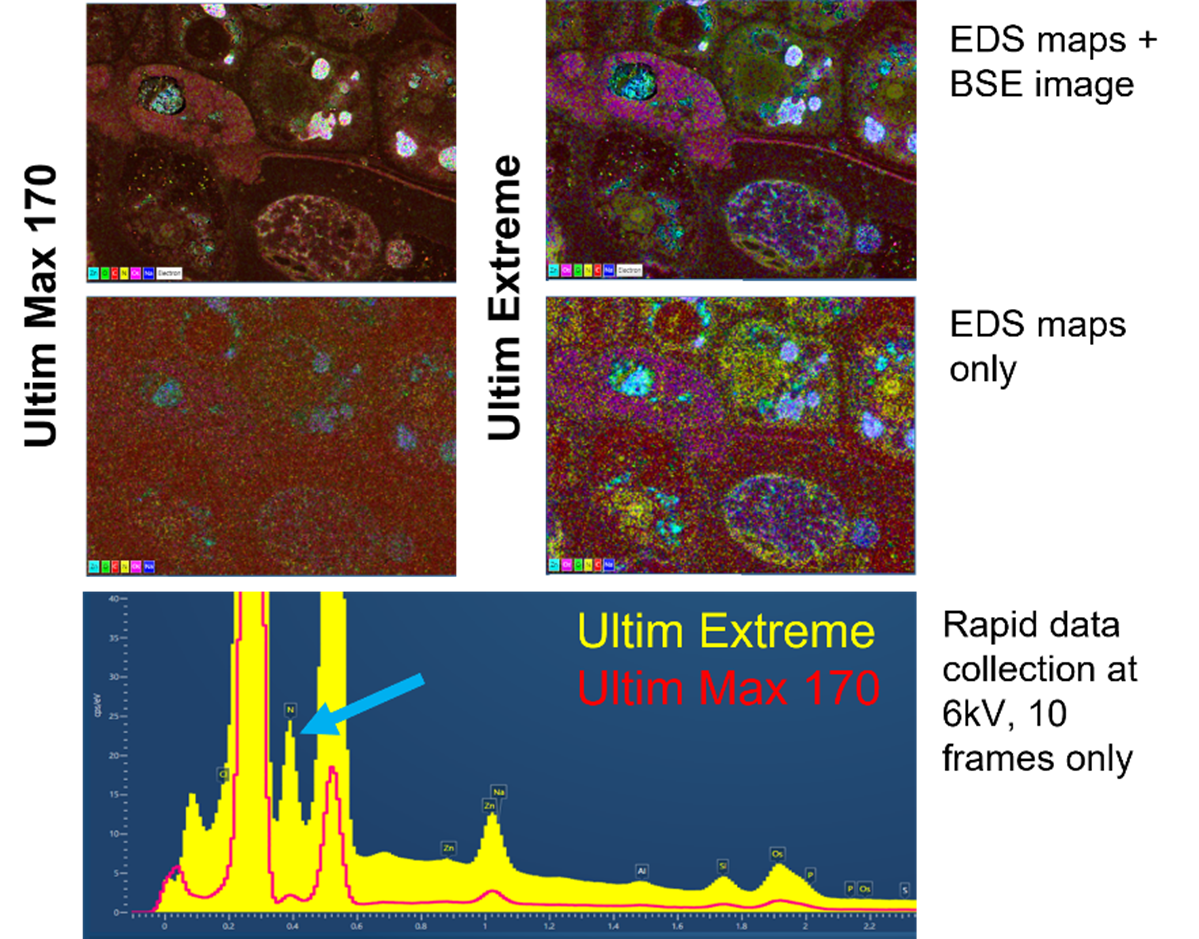
Figure: A comparison between the Ultim Extreme detector and the Ultim Max 170 showing maps and spectra taken of a resin embedded Venus’ fly trap leaf and gland. Note the improved signal in the Ultim Extreme maps and in particular the yellow signal, which is nitrogen. There is a far more obvious nitrogen peak (blue arrow) in the Ultim Extreme spectrum (yellow) compared to the Ultim Max 170 (red).
Traditional SEM EDS uses relatively high accelerating voltages (20-30 kV) and high beam currents to produce strong x-ray emissions from samples. Biological SEM uses low accelerating voltages and beam currents to prevent damage to the sample, reduce the chance of charging and to improve image resolution.
The other aspect to consider is that biological samples are mostly comprised of light elements (hydrogen, carbon, nitrogen, oxygen, sodium, magnesium, phosphorous, sulphur, chlorine, potassium and calcium) and some heavier elements that are often found in trace amounts (chromium, manganese, iron, cobalt, copper and zinc). This means that the x-rays emitted from the sample are relatively low energy. Low energy x-rays can be absorbed by the windows that most EDS detectors have, which reduces the amount of x-ray signal that can be detected.
Improving Sensitivity for Biological Samples
The Ultim Extreme detector is designed without a window and with a modified snout, which has several advantages.
- The detector is sensitive to very low energy x-rays that can be absorbed by windows. Nitrogen signals in particular are often affected.
- The modified snout means the detector is positioned closer to the pole-piece of the microscope. This has two advantages.
- The detector is closer to the sample and can collect more of the x-rays emitted by the sample.
- The optimised position of the sample is closer to the pole piece of the microscope, improving the electron image resolution.
- The Ultim Extreme has a large area sensor, improving the speed at which maps can be acquired. Reducing accumulated beam exposure on biological samples reduces drift and charging artefacts as well as reducing specimen damage.
- Sensitive electronics mean that even the relatively low x-ray emission from biological samples can be detected, mapped, and quantified.
- A combination of all the above means that the Ultim Extreme detector is 10-30x more sensitive than other large area detectors for low energy x-rays.
New Avenues of Research through EDS
Whichever detector you end up choosing, there is no doubt that EDS offers exciting new avenues for biological research. As highlighted in a previous blog, EDS can provide the following:
- Fast, compositional identification of unknown features within and on biological samples
- Coloured electron micrographs for improved interpretation of ultrastructure
- The relative quantities of native and non-native elements within samples.
The ability to combine ultrastructural analysis of samples with sample composition is applicable to all areas of biological electron microscopy, and the Ultim Extreme detector is an ideal system to address this line of research.
For more information about the Ultim Extreme detector, take a look at our webpages or watch our recent webinar on Advanced techniques in SEM and EDS for biological samples.
For more information about biological EDS, please take a look at our application pages and our library of previous blogs and webinars on this topic.


