2nd December 2020 | Author: Dr Louise Hughes
What is it? Where is it? How much? Using EDS for biomaterials research
What is it? Where is it? How much? These are all questions that can be readily answered by using EDS when studying biomaterials and their interactions with tissues and cells.
What is it?
One of the main jobs of EDS often involves the identification of an unknown feature, for example, the composition of exogenous material that has appeared in patient samples. Some interesting examples of this are presented in our upcoming webinar “Biomedical research using energy dispersive x-ray spectroscopy”, covering areas where identification of material in patient samples plays a key role in clinical research and in treating patients. I will also describe an example using implant derived wear particles found in patient tissue following implant failure in a previous blog. Identification of these types of features can be crucial in ensuring the best outcome for patients but also in understanding the mechanism behind exposure and how this may be addressed in the future.
The identification of materials is also important to ensure correct interpretation of data. In the example shown below, cobalt nanoparticles were fed to macrophages to analyse uptake and potential cytotoxicity (fig. 1). In the backscattered images we can clearly see clusters of dense particles that could be assumed to be the material we are interested in due to their density. However, EDS analysis shows that some of these are composed of material that is likely derived from the cell culture techniques and are not all cobalt nanoparticle clusters (fig 1). It is important that we can characterise our materials correctly. The BSE signals also did not detect nanoparticles from deeper within the cells, but they were detected using EDS.
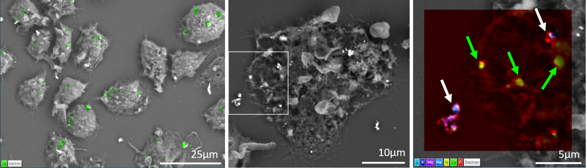
Fig. 1. Images showing backscattered electron data of macrophage cells that have been fed cobalt nanoparticles. Left image shows an EDS map overlay of cobalt (green). Right image shows a layered EDS map with cobalt (green colour and green arrows), and combined signals from silicon (light blue), potassium (dark blue), magnesium (purple) and sodium (medium blue) (white arrows) with carbon (red) and nitrogen (yellow) signals in the background. The cobalt provides backscattered contrast, as can be seen in the images, but other material may also provide a similar signal and EDS was necessary to correctly identify the cobalt signals. Sample was provided by Rachel Vowden and Zhidao Xia (Swansea University).
Where is it?
Many electron microscope research questions revolve around the localisation of features of interest, which can be achieved by the application of specific stains, labels, ultrastructural analysis, and CLEM, amongst others. EDS, applied as an imaging tool to produce colour electron microscopy based on elemental composition, offers an alternative to these options. EDS was used to examine the biodegradation of a hydroxyapatite sample that was not suitable for either TEM or confocal microscopy. The application of stains was not enough to provide sufficient differentiating contrast to positively identify boundaries between cells and the biomaterial. EDS provided information that enabled correct interpretation of image data, providing elemental contrast that supplemented and enhanced the electron image information (fig. 2)
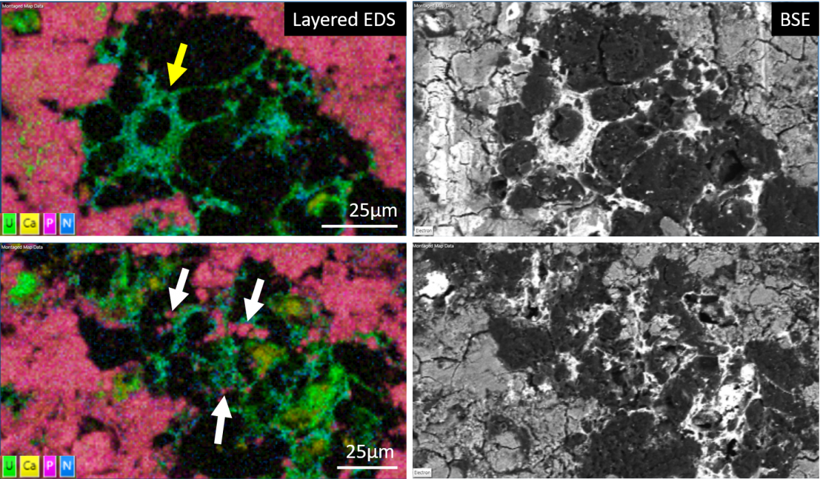
Fig. 2. Images show layered EDS maps (left) and backscattered electron images (right) showing cells interacting with a biodegrading hydroxyapatite implant. The top images show a distinct stellate cell (yellow arrow), but aside from this once cell, it proved difficult to identify the difference between cells and the biomaterial elsewhere. EDS not only provides clear contrast between the cells and surrounding material but also enabled easy identification of features such as small piece of implant that had broken off and were surrounded by cells (white arrows), providing valuable information about the degradation process. . Sample was provided Zhidao Xia (Swansea University).
How much?
As well as localising material, EDS provides a rapid method for characterising and measuring the composition of your biomaterials at any stage of manufacturing and failure analysis, as well as following in vivo and in vitro testing. The advantage is that you can combine your ultrastructural analysis with determination of material composition, maximising productivity in a rapid, easy, and non-destructive (for materials) technique. For more information about quantitative analysis take a look at our series of blogs by Simon Burgess (What is standardless quantitative analysis) (Be confident in your EDS results and maximise your productivity).
You may also interested in our on-demand webinar about “Operating SEM-EDS in a regulated environment” which has focus on common applications within the pharmaceutical industry.