Experimental Strategy & Results
We therefore collaborated with Oxford Instruments, Inc., Pleasanton, CA to test a novel backscattered electron and x‑ray (BEX) imaging technique using their “Unity” detector that is 8x more sensitive than the largest available EDX detectors. The “Unity” detector consists of very sensitive dual BSE and x‑ray sensors for the simultaneous and correlated detection of backscattered electrons (z‑contrast imaging of ultrastructural tissue morphology) and characteristic x‑ray signals (elemental analysis for metal detection and mapping) which results in unprecedented speeds of data acquisition and sensitivity due to its geometry and proximity. These characteristics suggest that the “Unity” BEX imaging system is suited for applications in ultrastructural immunohistochemistry. We therefore wanted to evaluate if detection, color coding and overlay of the x‑ray signals over BSE‑SEM morphology images would allow for the localization of metal‑labeled antigens associated with cells and organelles with nanometer resolution and precision in a timely manner.
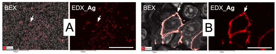
As a first experimental model system we used cryo‑sections of human tonsils that were single‑labeled with primary antibodies against CD11c, a molecular marker associated with the plasma membrane of dendritic cells and macrophages, and employed an enzymatic silver deposition reaction for secondary detection (Fig.1, A and B). In these images dendritic cells and macrophages are identified by specific silver precipitates associated with the cell membrane (Fig.1, A and B, arrow).
As a second experimental model system we used resin sections of mouse pancreatic tissue, that were single‑labeled with a specific primary antibody against the hormone insulin, followed by secondary detection with either 20 nm colloidal gold particles (Fig.1, C and D), or by a secondary detection using the enzymatic formation of silver particles (Fig.1, E and F). Therefore, in the pancreas sections the presence of insulin, which is packaged and transported in secretory vesicles, is revealed by elemental mapping of gold (Fig.1, C and D, arrowhead) or silver (Fig.1, E and F, circle).
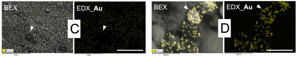
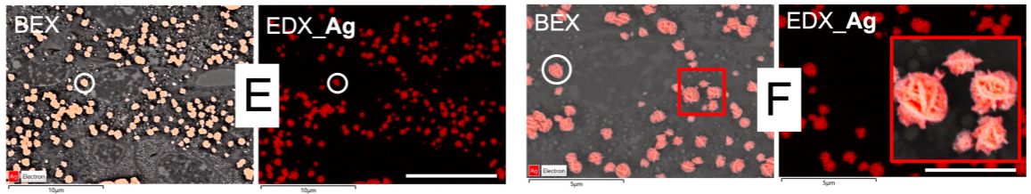
The results show that the “Unity” BEX imaging system can be efficiently applied for ultrastructural immuno‑localization of antigens by elemental mapping of either gold‑nanoparticles or silver‑precipitates. All images were acquired within a few seconds or minutes. This allowed for the efficient acquisition of overview maps of tissue sections to find labeled cells and structures of interest (Fig.1, A, C and E) before imaging at a higher magnification (Fig.1, B, D and F). The BEX technique unequivocally enabled the spatial detection of gold (EDX_Au) within the 20 nm sized colloidal gold particles used for anti‑insulin labeling (Fig.1, D, arrowhead) and, furthermore, revealed substructures in the enzymatically grown silver particles (Fig.1, F, inset, EDX_Ag). These promising results suggest that multicolor immuno‑EM strategies can be developed by combining multiplexed immunohistochemistry based on multiple heavy metals, followed by multiplexed elemental analysis and mapping with the “Unity” BEX imaging technique.

