Time travel with the Winchcombe meteorite EDS analysis
17th March 2022 | Author: Dr Haithem Mansour
Time travel with the Winchcombe meteorite EDS analysis
Analysing meteorite samples in an SEM is always exciting but analysing a piece of rare carbonaceous chondrite meteorite, estimated to be around 4.6 billion years old, is incredibly good fun! It is like time travelling!
Recently, I had the privilege to analyse the Winchcombe meteorite that fell in the little Gloucestershire town , in February 2021. The extra-terrestrial rock fell to earth in a spectacular meteor fireball event that was witnessed by over a thousand people in the UK. The UK Fireball Alliance (UKFAII) quickly responded to the event and with the help of local residents in Winchcombe, recovered about 600g of meteoritic material in an excellent state for scientific analysis. The fragments didn’t experience rainfall prior to collection and were placed in a protected environment within 12 hours from the fall, meaning that the quality of the samples is similar to those retrieved from asteroids by space probes.
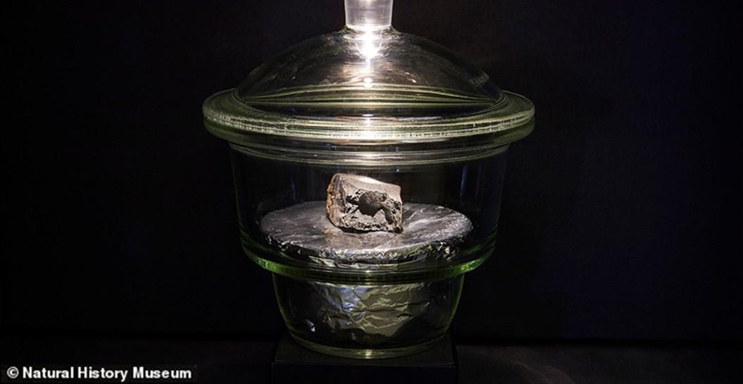
The largest part of the meteorite (figure 1) is now displayed in the Natural History Museum of London. Other parts were shared with different laboratories across the UK to do meticulous analysis with different techniques, forming the UK Winchcombe consortium study.
Figure 1: Winchcombe meteorite displayed at the Natural History Museum of London. Photograph from Trustees of the Natural History Museum, London
Scrutiny of the meteorite showed that it is a rare carbonaceous chondrite meteorite of similar age to the Earth itself, at just below 4.6 billion years. Understanding the microstructure and composition of this meteorite can give insights into how our planet formed and what the early solar system was like, which in turn can give important clues about the origins of the oceans and life on earth.
Oxford Instruments is part of the UK Winchcombe consortium study, and we received a sample of the meteorite from the Natural History Museum. In this blog, I will show a few preliminary results highlighting the magnificent microstructure of this meteorite that we are beginning to study.
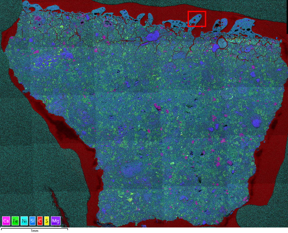
For this analysis, I used a high resolution FEG-SEM equipped with an Ultim Max 170 EDS detector and AZtecLive software. To have a good overview of the sample, I started by collecting a large area map of the whole sample, i.e. a series of simultaneous electron images and X-ray maps stitched to form one large map (Figure 2). It gave an initial overview of the chondrules and metal grains present. The meteorite sample is surrounded by conductive carbon resin in red as displayed in figure 2.
Figure 2: A series of individual maps stitched together to form a Large Area Map of the meteorite sample using AztecLive software. A coloured image combining X-ray element maps of the same region.
Using Live Chemical Imaging (LCI) gave me a great advantage to find areas of interest easily and quickly. Thanks to this new technology, it is possible to navigate and see the X-ray elemental maps live; a game changer compared to the conventional “move, stop, collect and repeat” workflow.
Here is a short video of the sample navigation using LCI:
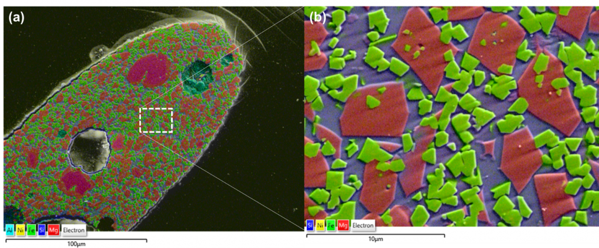
As shown in the video, LCI provides simultaneous electron imaging and X-ray mapping during navigation, which is very useful for locating areas with distinctive composition. In the video, I focused on the area located on the external top layer of the meteorite in figure 2, ranging from 0.5 µm to a few hundreds of microns in thickness. It clearly has a different structure to the rest of the sample. This surface layer is known as a meteorite fusion crust. A fusion crust is a thin melted layer transformed under high temperatures when the meteorite entered our atmosphere at very high speed (typically between 15 and 70 km/seconds). Although it takes only 4 to 5 seconds for the meteorite to pass through the atmosphere, the event generates so much energy it melts the surface of the meteorite (quite evident from the spectacular fireball seen by so many observers!).
Figure 3: (a) EDS map combined with secondary electron image from the fusion crust layer of the Winchcombe meteorite at 1,000X magnification and (b) at 10,000 X magnification
Figure 3 shows two high resolution maps of the fusion crust at different magnification collected from the area highlighted with a red rectangle in the large area map (figure 2). The analysis shows a fine-scaled microstructure with faceted iron oxide and olivine grains in a silicate-rich matrix. The core part of the meteorite has several fascinating microstructures, and can’t be shared in one blog, but I will just add another figure of one of the most beautiful EDS maps I collected.
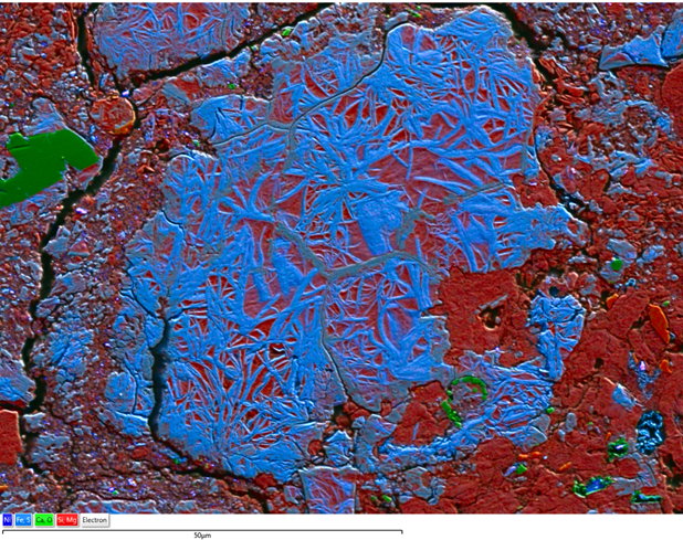
Figure 4: Ultra-high resolution EDS map collected using Ultim Max 170. Fe-rich serpentine veins structure occurring in an altered olivine grain
Figure 4 is an ultra-high resolution EDS map combined with an electron image. It shows an Fe-rich serpentine vein structure (in blue) occurring in an altered olivine grain (in red).
I hope you enjoyed this blog in which I gave a glimpse about the fascinating microstructure of the meteorite. I am an electron microscopist with a material science background but working on this sample allowed me to learn a good deal about meteorites. It is a very captivating subject.
Please keep an eye out for more posts on the Winchcombe meteorite as our research progresses!